
The Marvel of Prenatal Brain Development: From Conception to Birth and the Care to Take
The journey of the human brain begins long before a baby takes its first breath. From a microscopic layer of cells to a highly complex and flexible control centre, prenatal brain development is a rapid and incredibly intricate process.
The pregnancy period is a complex system and nature networked it systematically. Pregnancy is a period that demands discipline, peace, and mental and physical wellbeing. But in the modern age, working women face long daily commutes, office and home stress, cravings for junk food, delayed meals with large portions, infection exposures, and exposure to stress or violence. All of these factors can impact the developing prenatal brain. In the present day, the rise of neurodiverse children with hyperactivity, speech and language delays, and sensory processing challenges can often trace back to these early environmental stressors.
Let us look at how the brain structurally and cellularly develops throughout the fetal stage:
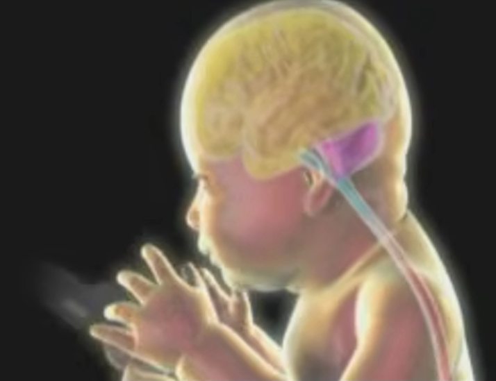
The Foundation: The First Four Weeks
In the first four weeks after conception, the central nervous system begins as a simple sheet of tissue in the outermost layer of embryonic cells called the neural plate.
- Forming the Tube: The neural plate folds to create a neural groove, which then curls into a closed structure known as the neural tube.
- The Four Key Regions: This tube rapidly differentiates into four crucial parts: the forebrain, midbrain, hindbrain, and spinal cord.
- Division of Labor: The forebrain will become the cerebral cortex (controlling sensory translation, complex thoughts, and memory). The midbrain becomes a relay station sending information across the brain. The hindbrain manages basic survival processes like heart rate and breathing, while the spinal cord acts as the information highway to the rest of the body.
Structural Expansion: Weeks 4 through 40
As the embryo grows rapidly, the physical shape of the brain and head transforms:
- Early Milestones (Weeks 4-8): The face becomes recognizably human, the eyes migrate to the front of the head, and two distinct hemispheres of the cerebral cortex emerge. By week 8, the major structural foundations of the brain are already in place.
- The Wrinkling Effect (Weeks 28-40): Initially, the brain is relatively smooth. But starting around week 28, the cerebral cortex expands massively in surface area to fit inside the skull. It begins to fold and wrinkle, creating hills (gyri) and valleys (sulci). These folds are critical; they allow for a vast neural network that makes the human brain a precise and flexible processor of information.
Cellular Growth and Networking
While the physical structures form, a booming microscopic construction project is happening at the cellular level:
- Neurogenesis and Neural Migration: New nerve cells are born in the innermost layers of brain tissue and migrate outward to their final destinations. They are guided by fibers from glial cells, acting much like gymnasts climbing ropes. The brain grows in layers, similar to an onion, with newer cells traveling further outward than the older ones.
- Making Connections (Synaptogenesis): Once a nerve cell reaches its destination, it acquires specific characteristics and sprouts extensions: dendrites (antennas that receive signals) and an axon (a larger extension that sends signals).
- Bridging the Gap: A synapse is the tiny gap between the axon of one neuron and the dendrite of another. When an impulse reaches the end of an axon, chemicals called neurotransmitters are released, acting as messengers to cross the gap and deliver the signal to the next cell. The brain creates so many of these connections that the process is known as exuberant synaptogenesis.
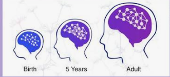
Synaptic Pruning: Sculpting Efficiency
To make the brain rapid and efficient, it eventually removes excess, unused synapses. This pruning process continues through infancy, childhood, and adolescence, effectively customizing the brain's circuitry in response to an individual's unique life experiences.
This incredible sequence of folding, migrating, connecting, and pruning ensures that by the time a baby is born, their brain is primed to learn, adapt, and make sense of the new world around them.